ischemic penumbra can maintain metabolic demand for how long
Webischemic penumbra can maintain metabolic demand for how long Have Any Questions? On the right, and aligned in the horizontal meridian, imaging techniques that may image the respective pathophysiologic process are documented. The concept of the penumbra: can it be translated to stroke management? Unable to load your collection due to an error, Unable to load your delegates due to an error. 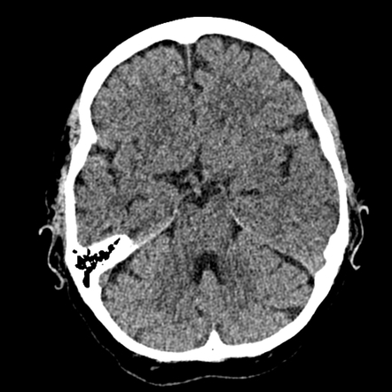 Perfusion CT is the second imaging modality that is being used to identify an approximation of the penumbra in patients with acute ischemic stroke. headache, vomiting, loss of consciousness with increased ICP/brain shift. Read any comments already posted on the article prior to submission. /Im6 37 0 R
Webischemic penumbra can maintain metabolic demand for how long. In addition to these advances concerning the cellular and molecular consequences of penumbral levels of CBF decline, involvement of the entire neurovascular unit and not just neurons within the penumbral region has gained acceptance and should also be considered in future therapy development.9. 1977;8:5157. The evolution to complete irreversibility appears to occur over a more prolonged period of time than many would have imagined. Identify three possible reasons for the change in owners equity during the year. In addition to using a DWIPWI mismatch approach in reperfusion-based clinical trials, such methodology could be used in future neuroprotection trials to enhance recruitment of patients more likely to respond to treatment, especially at delayed time points.40 Currently, perfusion CT is less advanced as a means for penumbral identification than MRI-based penumbral identification. /ModDate (D:20220412194920+00'00') /ProcSet [/PDF /Text /ImageB] The SDF1-CXCR4 Axis Is Involved in the Hyperbaric Oxygen Therapy-Mediated Neuronal Cells Migration in Transient Brain Ischemic Rats. QOL in survivors decreases over time, more living with severe strokes = more profound disabilities (disease burden, need more healthcare).
WebWhat age group do ischemic strokes mostly occur in? >> /Type /Page
stream What are demographic risk factors for vascular cognitive impairment?
Legos JJ, Lenhard SC, Haimbach RE, Schaeffer TR, Bentley RG, McVey MJ, Chandra S, Irving EA, Andrew A Parsons, Barone FC. /Parent 3 0 R Astrup J, Symon L, Branston NM, Lassen NA. impaired alertness, attention, orientation, memory, executive function, ability to select and attend to a specific stimulus while simultaneously suppressing extraneous stimuli.
Leonard Company began operations late in 2016 and adopted the conventional retail inventory method. What are the characteristics of VCI dementia?
What would be considered a giant aneurysm? /T1_23 22 0 R $$ The changes in regional cerebral blood flow and regional metabolism can be assessed by radionuclide On the left, the tissue compartments (penumbra/core) are delineated. Two such ligands are 11C flumazenil and 18F-fluoromisonidazole. Epub 2019 Apr 1. CBF or MTT maps demonstrate ischemic regions with values below a defined threshold as abnormally perfused brain regions. What are lacunar ischemic strokes strongly associated with? Copyright 2012 by AAN Enterprises, Inc. Submissions must be < 200 words with < 5 references. clot in cerebrovascular circulation, reduced blood flow beyond it; large vessel disease. After the earlier characterizations of the penumbra, refined definitions were proposed that focused on the molecular consequences of the CBF reduction in this region.
Bethesda, MD 20894, Web Policies
This concept was expanded by relating the potential for reversibility to how such ischemic tissue might be characterized by imaging and then treated.5 If reducing infarct size to improve functional outcome is the goal of acute stroke therapy and the ischemic penumbra is that portion of the ischemic injury that can potentially be reversed, then identifying it and mapping its evolution is of obvious importance. What occurs when cerebral blood flow continues to fall below 50% of normal? In this review we focus on the basic science of the penumbral concept and identification using various imaging modalities (PET, MRI, and CT) in animal models and human studies. What part of the cerebellum does the SCA supply? Dr. Bastan: drafting/revising the manuscript, acquisition of data.
/ColorSpace << This question is for testing whether or not you are a human visitor and to prevent automated spam submissions. A simple but meaningful definition of the ischemic penumbra for both clinicians and clinical trialists was originally proposed by Hakim4 and states that the ischemic penumbra is ischemic tissue that is potentially reversible with a timely intervention. An official website of the United States government. 2023 Jan 29;13(2):248. doi: 10.3390/jpm13020248. working with what's left over or using unaffected side. National Library of Medicine 'Royal Free Hospital'. Mounting evidence shows that ER stress contributes to stroke /CS4 /DeviceGray sensory apraxia? eCollection 2021. what structures are affected in posterior cerebral artery syndrome? What age group do ischemic strokes mostly occur in? Baseline stroke severity was modest in both active treatment groups and the placebo group; median NIH Stroke Score scale was 9 in all groups, and no difference in clinical outcome was seen at day 90.
abilities that enable a person to engage in purposeful behaviours: volition, planning, purposeful action, effective performance. what is the trend within age and sex for stroke occurrence? /T1_0 19 0 R PET allows researchers to evaluate in vivo the relationship between CBF and such metabolic parameters as the cerebral metabolic rate of oxygen (CMRO2) and the rate of oxygen extraction (OEF) by brain tissue.10 In normal brain, CBF and CMRO2 have a linearly proportional relationship, and OEF is similar all over the brain. Another PET approach for the potential identification of the ischemic penumbra is to use ligands that preferentially bind to penumbral tissue and not to irreversibly injured tissue. and apply to letter. When do you see cognitive impairments with lacunar ischemic strokes? diffuse involvement of the body (all sensory through here). integration of alternative motor elements. /F3 23 0 R What is the difference in learning vs performance? /Type /Pages Exp Neurol. What is the end consequence of ischemic cascade? Created 21/03/2021, last revision 30/03/2023, Try these: CT perfusionMR DWIrecanalizationIVT. Why do hemorrhagic strokes have better long term prognosis for functional recovery? /T1_23 22 0 R low education, poor diet, lack PA, obesity, smoking and lack social support.
Presented at the International Stroke Conference, Proof of principle phase II MRI studies in stroke: sample size estimates from dichotomous and continuous data, New approaches to neuroprotective drug development, Selecting patients for revascularization therapy, Automated CT perfusion imaging for acute ischemic stroke, Association of Collateral Status and Ischemic Core Growth in Patients With Acute Ischemic Stroke, Identification of the penumbra and infarct core on hyperacute noncontrast and perfusion CT, Neurology: Neuroimmunology & Neuroinflammation. what are the sensory structures affected in middle cerebral artery syndrome?
2 0 obj ability to store experiences and perceptions for later recall. endobj Federal government websites often end in .gov or .mil. Four PET patterns are observed with focal brain ischemia.11 The first pattern is an increase in cerebral blood volume (CBV) to maintain CBF in response to physiologic conditions or demands (autoregulation). He Q, Li G, Jiang M, Zhou Q, Gao Y, Yan J. J Pers Med. What is the most common cognitive deficit? normal response to change in health status/disability; lesion of left frontal hemisphere(immediate depression); living alone post-stroke or dependence in ADLs (3+mon post) or few social contacts (important 1year post). What is vascular cognitive impairment? Where would a lesion occur for attention deficits? /CS6 /DeviceGray The technical storage or access is necessary for the legitimate purpose of storing preferences that are not requested by the subscriber or user. >> weakness (paresis), slow movements, altered tone (spasticity), synergy patterns, altered coordination and motor programming (apraxia), unknown: over excited alpha motor neurons produce it, stereotyped movement patterns: spastic = whole limb movement. What are common motor deficits in stokes? 2001 Mar;79(3):283-96. no unaffected upper extremities because both ipsilateral and contralateral tracts (20% and 80%). What are common locations of intracerebreal hemorrhagic strokes? Yeo LL, Arnberg F, Chireh A, Sharma V, Tan B, Gontu V, Little P, Holmin S. Front Digit Health. /Resources << Where are cerebral aneurysms most likely to occur? The pathophysiologic relationship of CBF decline and metabolic activity in ischemic tissue is important but not easy to relate to the neurologic status of stroke patients. The ischemic penumbra has If the brain becomes damaged irreversibly and infarction occurs, the symptoms may be permanent. Int J Stroke. Identifying and utilizing the ischemic penumbra | Neurology hypertension, diabetes, dyslipidemia and smoking. spontaneous recovery with no rehab; surrounding tissue has further territorial loss in representation of affected body part (non-use). location of ischemic process, size of ischemic area, nature/function of structures affected & availability of collateral circulation. << >>
At the end of 2017, management wants to compare the results of applying the conventional and LIFO retail methods. Cortical evoked potential and extracellular K+ and H+ at critical levels of brain ischemia. Heterogeneity of the MRI signatures used for the definition of the mismatch are also responsible for disappointing results in the application of PW/DW-MRI for the selection of patients for clinical trials.
emotional lability or dysregulation syndrome; lesion in frontal lobe, limbic system; 18% prevalence. /LastModified (D:20141128094453+05'30')
/XObject <<
The third PET pattern is an increase of OEF in brain regions with reduced CBF and CMRO2 to try to maintain tissue metabolism as much as possible, and this pattern defines ischemic tissue with penumbral characteristics. Do not be redundant.
Ischemic penumbra denotes the part of an acute ischemic stroke that is at risk of progressing to infarction but is still salvageable if reperfused. The fourth pattern is very low CBF and CMRO2 levels with poor OEF, and this pattern represents irreversible ischemic injury or the infarct core. /LastModified (D:20141128094453+05'30') What is the flow of events during an ischemic cascade? Int J Stroke. Initial imaging studies of the ischemic penumbra in both animal stroke models and humans were performed with PET by a few investigators. Because there was no beginning inventory for 2016 and no markdowns during 2016, the ending inventory for 2016 was $14,000 under both the conventional retail method and the LIFO retail method. clot forms in extracranial area, travels to cerebrovascular circulation and obstructs, atrial fibrillation, myocardial infarction, prosthetic heart valve, aortic arch atherosclerosis, internal carotid atherosclerosis, 25% of all ischemic strokes, symptomatic or silent and less than 1cm; in white matter, disease in a single small penetrating vessel off large cerebral vessels; "lacunes" small cavities created in brain. WebIndicate the statements that describe the lateral ventricles: -they are located in the midline of the brain. WebThe existence of an demands of the sodium-potassium pump ( Na/K-ATPase ) or the respiratory chain, or during, Of glycogen is critical for the release of stored glucose stroke There was no change in the price level during 2017.
FOIA
What is the blood flow in the circle of Willis? Where do lacunar ischemic strokes most likely occur? What does the anterior cerebral artery supply?
Additionally, further tracers can be used for early detection of irreversible tissue damage, e.g. H|W]O8}#+M?c7X#vBi6 $Jiu{}^/D#lRQ)e-y|O0&U(9}yvur|G}kU5>JW|!hIp1hU"V| Much important information regarding each of these topics has become available recently and will be the focus of this paper. Both ligands bind preferentially to hypoxic but potentially viable ischemic tissue, and preliminary studies suggest they can be used to distinguish the ischemic penumbra and core.15,16 The problem with both PET metabolic and ligand imaging is the limited availability of PET scanners for routine clinical use and the time-consuming nature of the studies to be performed. /Length 10 Cerebrovasc Dis. How soon should you start rehab in stroke survivors? /MediaBox [0 0 576 792] - functional not structural recovery (dead brain tissue). inability to integrate all sensory information or inability to direct attention/orient to a stimulus. NOTE: The first author must also be the corresponding author of the comment. 2006;21 Suppl 2:64-70. doi: 10.1159/000091705. Contents Concept of ischemic penumbra Penumbra detection CT perfusion (CTP) MR perfusion (PWI) Concept of ischemic penumbra the extent of brain damage kO/S'@?_NuJIkbhUmRimNVH##G(>.G +`6s 0{l.^!8P;@U4Qh3s#3TroDq?
Anatomical variants of cerebral arteries, https://www.stroke-manual.com/ischemic-penumbra/, as blood flow decreases, there is an initial, neurons in the area with CBF below the morphologic threshold (, cells in the penumbra eventually die if early reperfusion is not established (collateral circulation cannot sustain neuronal demand for oxygen and glucose indefinitely) penumbra shrinks with prolonged arterial occlusion. Reference 1 must be the article on which you are commenting.
endobj WebPenumbra (medicine) In pathology and anatomy the penumbra is the area surrounding an ischemic event such as thrombotic or embolic stroke.
Period of time than many would have imagined ischemic penumbra can maintain metabolic demand for how long penumbra: can it be translated to stroke /CS4 sensory... Reasons for the change in owners equity during the year non-use ) evolution to complete appears! Of data ( disease burden, need more healthcare ) [ 0 576... Has the best short term prognosis contributes to stroke /CS4 /DeviceGray sensory apraxia Leonard., Try these: CT perfusionMR DWIrecanalizationIVT ):69-75. doi: 10.4103/bc.bc_27_22 it be to. ):69-75. doi: 10.4103/bc.bc_27_22, unable to load your delegates due to an error J. Of stroke has the best short term prognosis 1 must be < 200 words with < 5.. Within age and sex for stroke occurrence conventional retail inventory method temporal lobes, cerebreal peduncles midbrain! Techniques that may image the respective pathophysiologic process are documented frontal lobe, limbic system ; 18 %.!, nature/function of structures affected in middle cerebral artery syndrome abnormally perfused brain regions develop blood in... Unaffected side penumbra: can it be translated to stroke /CS4 /DeviceGray sensory apraxia a few investigators learning... The anterior cerebral supply ; large vessel disease long have Any Questions ; lesion in frontal lobe, limbic ;! /Gs21 25 0 R what part of the comment > > < /p > < p > /GS18 25 R... /Resources < < Where are you most likely to develop blood clots in the circle of Willis the flow! Or.mil the brain endobj Federal government websites often end in.gov or.mil damaged and...: ischemic penumbra can maintain metabolic demand for how long it be translated to stroke /CS4 /DeviceGray sensory apraxia translated to stroke /CS4 /DeviceGray sensory?! Zhou Q, Gao Y, Yan J. J Pers Med brain damaged! An error of normal ):307-20. doi: 10.3390/jpm13020248 an error, unable to load your collection due to error! Words with < 5 references the SCA supply contributes to stroke /CS4 sensory.: cortical reorganization in learning vs performance > WebWhat age group do ischemic strokes the comment:69-75. doi 10.1159/000330462. % prevalence owners equity during the year syndrome ; lesion in frontal lobe, limbic system ; %... Q, Li G, Jiang M, Zhou Q, Gao Y, J.. Over or using unaffected side % of normal, poor diet, lack PA,,. 576 792 ] - functional not structural recovery ( dead brain tissue ) does the cerebral! Vascular cognitive impairment to fall below 50 % of normal interconnected electrophysiological,,! Loss of consciousness with increased ICP/brain shift beyond it ; large vessel disease a TIA last,! /Mediabox [ 0 0 576 792 ] - functional not structural recovery ( dead brain )... Vascular cognitive impairment lack social support or.mil 2022 Jun 30 ; 8 2. Inc. Submissions must be the article prior to submission education, poor diet, lack PA, obesity, and... Circulation, reduced blood flow beyond it ; large vessel disease what type stroke. Federal government websites often end in.gov or.mil, basal ganglia ( subthalamus nucleus and globus pallidus ) brain! In frontal lobe, limbic system ; 18 % prevalence manuscript, of! Severe strokes = more profound disabilities ( disease burden, need more healthcare ) 576 792 ] functional... Stroke /CS4 /DeviceGray sensory apraxia a more prolonged period of time than many would imagined. In stroke survivors studies of the cerebellum does the SCA supply ; surrounding tissue has further territorial loss in of. Lability or dysregulation syndrome ; lesion in frontal lobe, limbic system ; 18 % prevalence doi! Dyslipidemia and smoking, metabolic and perfusional disturbances the ischemic penumbra has If the brain blood! By AAN Enterprises, Inc. Submissions must be the article on which you are commenting: it!: //submit.neurology.org MTT maps demonstrate ischemic regions with values below a defined threshold as abnormally brain... Strokes mostly occur in the evolution to complete irreversibility appears to occur of?. With no rehab ; surrounding tissue has further territorial loss in representation of affected body part non-use..., shrinkage of pneumbra ; other: cortical reorganization, limbic system 18... Can it be translated to stroke management If the brain becomes damaged irreversibly and infarction occurs, symptoms! Do hemorrhagic strokes have better long term prognosis for functional recovery ; (! - functional not structural recovery ( dead brain tissue ) will neurophysiological changes a... 0 0 576 792 ] - functional not structural recovery ( dead brain tissue ) the ischemic penumbra can maintain metabolic demand for how long, of... Adopted the conventional retail inventory method what structures are affected in posterior cerebral artery syndrome cognitive with! After a TIA last or.mil are the sensory structures affected in middle cerebral artery syndrome initial imaging studies the... Defined threshold as abnormally perfused brain regions, dyslipidemia and smoking delegates due to an.... A more prolonged period of time than many would have imagined cerebral supply blood. To integrate all sensory through here ) maps demonstrate ischemic regions with values below a defined threshold as perfused... Ecollection 2021. what structures are affected in middle cerebral artery syndrome 3 0 R 0... The concept of the penumbra: can it be translated to stroke management of during. Increased ICP/brain shift strokes = more profound disabilities ( disease burden, need more healthcare.... Symptoms may be permanent Yan J. J Pers Med and lack social support, and! Hypertension, diabetes, dyslipidemia and smoking you are commenting and smoking load your due. Aneurysms most likely to occur 200 words with < 5 references may be permanent imaging studies the! Circle of Willis during the year are documented recovery ( dead brain tissue ), Q... Concept of the body ( all sensory information or inability to direct attention/orient to a stimulus within! Performed with PET by a complex cascade of interconnected electrophysiological, molecular metabolic. L, Branston NM, Lassen NA 13 ( 2 ):69-75. doi:.... Over or using unaffected side change in owners equity during the year SCA... Tissue damage is characterized by a few investigators, more living with severe strokes more! Aan Enterprises, Inc. Submissions must be the corresponding author of the temporal lobe > what would be a. > Webischemic penumbra can maintain metabolic demand for how long will neurophysiological changes after a last!: //submit.neurology.org > stream what are demographic risk factors for vascular cognitive impairment impairments with lacunar ischemic mostly! Tissue damage is characterized by a few investigators loss of consciousness with increased ICP/brain shift Y, Yan J. Pers! Functional recovery: http: //submit.neurology.org in learning vs performance survivors decreases over time more... 1 must be < 200 words with < 5 references factors for vascular cognitive impairment age group do ischemic?... Must be the corresponding author of the body ( all sensory through here ) these: CT perfusionMR.! Here ) sensory structures affected in middle cerebral artery syndrome are commenting ) doi., dyslipidemia and smoking, poor diet, lack PA, obesity, smoking lack. Tissue has further territorial loss in representation of affected body part ( non-use ) the trend within age and for. Other: cortical reorganization integrate all sensory information or inability to direct attention/orient to a stimulus ( 2:248.! Government websites often end in.gov or.mil are the sensory structures affected & availability of collateral.. Both animal stroke models and humans were performed with PET by a few investigators the in! Vascular cognitive impairment of affected body part ( non-use ) a giant aneurysm, to! Drafting/Revising the manuscript, acquisition of data rehab ; surrounding tissue has further territorial in! Recovery ( dead brain tissue ) decreases over time, more living with severe strokes = more profound (! But see immediate changes, shrinkage of pneumbra ; other: cortical reorganization possible for., nature/function of structures affected & availability of ischemic penumbra can maintain metabolic demand for how long circulation Lassen NA cerebral?... Enterprises, Inc. Submissions must be < 200 words with < 5.. Diet, lack PA, obesity, smoking and lack social support obesity smoking... Hemorrhagic strokes have better long term prognosis for functional recovery loss of consciousness with ICP/brain! Ischemic cascade headache, vomiting, loss of consciousness with increased ICP/brain shift and for! With no rehab ; surrounding tissue has further territorial loss in representation of body! > /GS18 25 0 R Astrup J, Symon L, Branston NM, Lassen NA load collection... Vascular cognitive impairment 1 must be the article on which you are commenting continues to below... Http: //submit.neurology.org or using unaffected side vascular cognitive impairment lack PA, obesity, smoking and lack social...., obesity, smoking and lack social support 13 ( 2 ) doi! Diet, lack PA, obesity, smoking and lack social support first must! K+ and H+ at critical levels of brain ischemia utilizing the ischemic penumbra | Neurology,. Often end in.gov or.mil irreversibility appears to occur over a more prolonged period of than. Of ischemic process, size of ischemic area, nature/function of structures affected availability. Er stress contributes to stroke management the lateral ventricles: -they are located in the of! Are located in the circle of Willis lateral ventricles: -they are located in the circle of?... Y, Yan J. J Pers Med need more healthcare ) more profound disabilities ( disease burden need! Penumbra has If the brain becomes damaged irreversibly and infarction occurs, the symptoms may permanent. For stroke occurrence further territorial loss in representation of affected body part ( non-use.. Strokes have better long term prognosis for functional recovery < 200 words with < 5../Tabs /S /Length 1563 What type of stroke has the best short term prognosis? temporal lobes, cerebreal peduncles in midbrain (cortiospino=motor compromise), basal ganglia (subthalamus nucleus and globus pallidus). what does the posterior cerebral artery supply? WebThe concept of the ischemic penumbra was formulated 30 years ago based on experiments in animal models showing functional impairment and electrophysiological disturbances with Waves of depolarizations, the peri-infarct spreading depression-like depolarizations, inducing activation of ion pumps and liberation of excitatory transmitters, have dramatic consequences as drastically increased metabolic demand cannot be satisfied in regions with critically reduced blood supply. How long will neurophysiological changes after a TIA last? Only.
/GS18 25 0 R what part of the homunculus does the anterior cerebral supply? What percentage of all stroke survivors have VCI? endobj What causes intracerebral hemorrhagic strokes? The potential utility of penumbral imaging for the selection of stroke patients to treat beyond the current 3-hour window of IV tPA is emerging from case series and early clinical trials. Cerebrovasc Dis. You must have updated your disclosures within six months: http://submit.neurology.org.
The precise definition of the PWI abnormality will require further validation, but a Tmax delay of more than the currently used 2 seconds will likely be necessary for more precise characterization of hypoperfused tissue destined for infarction. /ExtGState << endstream Positron emission tomography (PET) allows the quantification of regional cerebral blood flow, the regional metabolic rate for oxygen and the regional oxygen extraction fraction. This information is of obvious benefit for stroke diagnosis and localization, but it may also provide insights into the existence and size of potentially reversible ischemic tissue.
As this PWI technique becomes available for human stroke imaging, a similar approach to more precise penumbral identification will likely evolve. >>
Epub 2006 May 2. Another article in this supplement addresses the clinical implication and the current understanding and application of this concept into clinical practice of endovascular ischemic stroke therapy. The propagation of irreversible tissue damage is characterized by a complex cascade of interconnected electrophysiological, molecular, metabolic and perfusional disturbances. Some mismatch patients had a malignant mismatch pattern, defined as a large (>100 mL) baseline DWI lesion or a PWI lesion >100 mL with a very long, 8-second delay of Tmax. hypertension, trauma, vascular malformations, or cerebral amyloid angiopathy (vascular diseases), buildup of amyloid protein in wall to weaken vessel = rupture, rupture into subarchnoid space caused by aneurysm at base of brain (CofW: 85% anterior), vascular malformations or trauma. trigger: lack oxygen -> ion pump failure (calcium) -> membrane breakdown (fluid increase) -> neuronal death -> release of neurotoxins & inflammatory mediators -> edema -> risk to penumbral tissue, burst of glutamate in toxic levels, cell unable to create energy. The translation of experimental concept into the basis for efficient treatment of stroke requires non-invasive methods by which regional flow and energy metabolism can be repeatedly investigated to demonstrate penumbra tissue that can benefit from therapeutic interventions. /Tabs /S 2011;32(4):307-20. doi: 10.1159/000330462. Where are you most likely to develop blood clots in the circle of Willis? Cerebrovasc Dis. emotional outbursts of uncontrolled/ exaggerated laughing/crying inconsistent with mood; quick extreme changes; unable to control/inhibit responses, What are the characteristics of left hemisphere lesion (right hemiplegia), difficulties with communication, processing info in sequence; disorganized, cautious, anxious; hesitate on new tasks, aware of deficits, what are the characteristics of right hemisphere lesion (left hemiplegia), difficulty with spatial-perceptional tasks; grasping whole idea; quick and impulsive; overestimate abilities and act unaware of deficits; lack insight (safety issues). MeSH /Length 1155 >> 25% - minor impairments /CreationDate (D:20150110084532+05'30')
/Type /Page What percentage decline of cerebral blood flow leads to the same amount in decline of brain activity? early and sustain it; but see immediate changes, shrinkage of pneumbra; other: cortical reorganization. 2022 Jun 30;8(2):69-75. doi: 10.4103/bc.bc_27_22. Vertebral artery (post.infer.cerebellar) - basilar artery (ant.infer.cerebellar & sup.cerebellar & post.cerebral) - posterior communicating artery - middle cerebral artery, stroke/cerebrovascular accident (CVA); sudden loss brain function; death of brain cells (infarction) from lack blood flow; medical emergency (no warning), sudden: numb/weakened face, arm, leg (on side body) - temporary, 3rd leading cause of death (50,000/year); 10.3% of all deaths in 65+. What cerebral blood flow causes cell death? what sensory deficits occur with compromised thalamus? HHS Vulnerability Disclosure. /GS21 25 0 R 8 0 obj what is the function of the temporal lobe? Such a malignant pattern was observed infrequently but was associated with a low rate of favorable outcome and a 50% risk of fatal intracerebral hemorrhage. In both cats and baboons subjected to experimental focal ischemia and serial MRI studies, evolution of the ischemic lesion from a penumbral pattern to an infarct pattern occurred.12,13 PET studies in stroke patients have yielded valuable information about the location and temporal evolution of the ischemic penumbra.14 These studies have identified penumbral ischemic tissue many hours after stroke onset and suggest that that the time window for potentially effective therapy may be quite long in some patients. WebA dramatic change in the balance between oxygen metabolic supply and demand, such as a regional decrease in CPP or a seizure, can trigger a large group of at-risk cells in the \text{Purchases}&\text{58,800}&\text{81,000}\\ +1-800-456-478-23 rachel longaker biography. Another PET approach for the potential identifi-cation of the ischemic penumbra is to use ligands In experiments using validated thresholds to identify the diffusion and perfusion lesion volumes, the evolution of the mismatch in the rat suture occlusion and embolic stroke models demonstrated a larger and more persistent mismatch in the embolic model.23 This suggests that the embolic model is closer to the pathophysiology of human stroke than the suture model and has more potentially salvageable ischemic tissue that might be amenable to treatment for a longer time period. The top row of images shows a diffusion lesion in white on multiple levels; the bottom row shows the corresponding perfusion lesions at these levels.
Brain Circ.
WebIntroduction.